
Sinoatrial Node: The Heart's Natural Pacemaker
The sinoatrial node (SA node), also known as the sinus node or Keith–Flack node, is a specialized region of cardiac muscle that serves as the heart's natural pacemaker. Located in the upper back wall of the right atrium, this oval-shaped structure is responsible for initiating the electrical impulses that drive the rhythmic contraction of the heart, establishing what is known as the sinus rhythm.
In a healthy heart, the SA node continuously generates electrical impulses called cardiac action potentials. These signals travel through the heart's electrical conduction system, triggering the muscle cells to contract and pump blood throughout the body. The rate of these impulses is dynamically adjusted by the nervous system to meet the body's oxygen demands.
Key Facts
- Function: Acts as the primary pacemaker by initiating cardiac action potentials.
- Location: Situated in the subepicardial layer of the right atrium, near the entrance of the superior vena cava.
- Dimensions: Approximately 10-20 mm long, 2-3 mm wide, and 1 mm thick.
- Blood Supply: Primarily supplied by the sinoatrial nodal artery.
- Control: Regulated by sympathetic (increases heart rate) and parasympathetic (decreases heart rate) nerves.
Anatomy and Structure
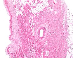
The SA node is a crescent-shaped structure embedded within the subepicardial layer of the right atrium. Its widest point is at the superior "head," tapering as it follows the sulcus terminalis toward the inferior vena cava. Structurally, it consists of a network of pacemaker cells encased in a dense matrix of connective tissue, which tends to increase in density as a person ages.
At a microscopic level, the node is a complex mesh of connective tissue containing collagen, fat, blood vessels, and nerves. The pacemaker cells themselves are smaller and paler than standard atrial cells, typically measuring about 8 micrometers in diameter and 20-30 micrometers in length. Because they contain fewer mitochondria, myofibers, and a smaller sarcoplasmic reticulum, these cells are less equipped for physical contraction than the cells of the atria or ventricles.
To ensure the SA node functions independently, it is insulated from the rest of the atrium by paranodal cells and connective tissue. Furthermore, the gap junctions (pores made of proteins called connexins) that allow electrical signals to pass between cells are smaller and fewer in number within the SA node, further isolating its activity from the surrounding atrial tissue.
Cellular Composition
The node is not uniform but contains a distribution of specific cell types:
- Central Area: A uniform mix of cardiac pacemaker cells, atrioventricular (AV) node cells, and Purkinje fibers.
- Crista Terminalis Region: Predominantly atrial cells (63±18%) and elongated spindle nodal cells.
- Septal Area: Mostly atrial cells (88±19%), with a presence of spider cells, spindle cells, and elongated spindle-shaped cells.
Blood Supply and Variation
The SA node is supplied by the sinoatrial nodal artery. This blood supply varies significantly between individuals. While most humans have a single artery, some may have two or three. Most commonly, this artery branches from the right coronary artery, though in some cases, it originates from the circumflex artery (a branch of the left coronary artery). Additionally, the artery may pass either in front of or behind the superior vena cava.
Physiology and Pacemaking
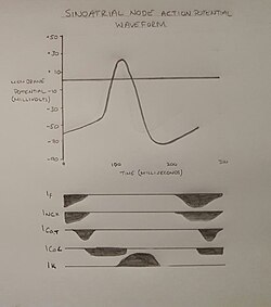
The primary role of the SA node is to initiate the action potential—a rapid change in membrane potential caused by the movement of ions. Unlike most cardiac cells, which have a stable resting potential, pacemaker cells exhibit a pacemaker potential. This means that immediately after repolarization, the cell automatically begins to depolarize again.
Once this automatic depolarization reaches a specific threshold potential, a full action potential is triggered. While other parts of the heart, such as the AV node and Purkinje fibers, can also initiate impulses, the SA node typically does so at a faster rate, allowing it to override other potential pacemakers.
Phases of the Action Potential
The pacemaker action potential is simplified into three main phases:
- Phase 4: The automatic depolarization (pacemaker potential) that leads toward the threshold.
- Phase 0: The rapid depolarization phase. When the threshold (-20 to -50 mV) is reached, L-type calcium channels open, allowing calcium (Ca) to flow into the cell.
- Phase 3: The repolarization phase, where the cell returns to a negative potential.
Neural Regulation of Heart Rate
The autonomic nervous system modulates the heart rate through two opposing pathways:
Sympathetic Stimulation (Positive Chronotropy)
Originating in the thoracic spinal cord (T1-T4), sympathetic nerves release noradrenaline. This binds to beta-1adrenoceptors, activating a Gs-protein and the cAMP pathway. The resulting cyclic adenosinemonophosphate (cAMP) binds to HCN channels, increasing the flow of sodium (Na) and potassium (K) into the cell. This accelerates the pacemaker potential, increasing the heart rate.
Parasympathetic Stimulation (Negative Chronotropy)
The Vagus nerves, originating in the brain, release acetylcholine. This binds to M2 muscarinic receptors, activating an inhibitory Gi-protein that blocks the cAMP pathway. Simultaneously, it activates GIRK-1 and GIRK-4 potassium channels, allowing K to flow out of the cell. This makes the membrane potential more negative, slowing the pacemaker potential and decreasing the heart rate.
Clinical Significance and History
If the blood supply to the SA node is blocked—often due to coronary artery disease or a myocardial infarction—the resulting ischemia can lead to cell death and sinus node dysfunction. If the SA node fails or the impulse is blocked, secondary pacemaker cells lower in the conduction system will take over to maintain a heartbeat.
The SA node was discovered in 1907 by medical student Martin Flack and his mentor, Sir Arthur Keith. The discovery was made while studying the heart of a mole in a makeshift laboratory in Kent, England.
| Feature | Detail |
|---|---|
| Primary Function | Initiates cardiac action potentials (Natural Pacemaker) |
| Location | Right atrium, subepicardial layer |
| Key Ion Channels | L-type Ca, T-type Ca, HCN, GIRK |
| Sympathetic Effect | Positive chronotropy (Increased heart rate) |
| Parasympathetic Effect | Negative chronotropy (Decreased heart rate) |
| Blood Supply | Sinoatrial nodal artery |
Frequently Asked Questions
What happens if the SA node stops working?
If the SA node fails or its signal is blocked, other latent pacemaker cells located further down the conduction system, such as those in the atrioventricular node or Purkinje fibers, will take over the role of initiating heartbeats, although usually at a slower rate.
How does the SA node differ from regular heart muscle cells?
SA node cells are smaller, paler, and contain fewer mitochondria and myofibers than atrial or ventricular cells. Most importantly, they lack a stable resting potential, allowing them to depolarize automatically.
What is "pacemaker shift"?
Pacemaker shift occurs when the specific cell that initiates the fastest action potential within the SA node changes location. In some animals, a superior shift increases heart rate, while an inferior shift decreases it.
Which nerves control the speed of the SA node?
The heart rate is controlled by the autonomic nervous system: sympathetic nerves increase the rate via noradrenaline, and parasympathetic nerves (specifically the Vagus nerves) decrease the rate via acetylcholine.
What is the role of the sinoatrial nodal artery?
The sinoatrial nodal artery provides the essential blood supply to the SA node. Blockage of this artery can lead to ischemia and cell death, resulting in sinus node dysfunction.