
TNF Signaling: Mechanisms of Immune Response and Cell Fate
Tumor Necrosis Factor (TNF) serves as a central mediator of the body's innate immune response—the rapid, non-specific first line of defense against invading pathogens. By interacting with two primary receptors, TNFR1 and TNFR2, TNF acts as a molecular switch that can dictate whether a target cell survives, proliferates, or undergoes programmed death.
While TNF typically promotes cell survival and inflammation by default, specific triggers—such as pathogen interference or co-stimulation with other cytokines—can shift the balance toward cell death. This versatility allows the immune system to either coordinate a systemic inflammatory response or eliminate infected cells to prevent viral or bacterial replication.
Key Facts
- Dual Receptors: TNF signals through TNFR1 (found in most cells) and TNFR2 (limited to specific cell types like endothelial cells and neurons).
- Cell Fate: Signaling can lead to survival (proliferation/inflammation), apoptosis (controlled death), or necroptosis (inflammatory death).
- TNFR1 Death Domain: Only TNFR1 possesses a cytoplasmic death domain, allowing it to directly trigger cell death.
- Reverse Signaling: Transmembrane TNF (tmTNF) can act as a receptor, triggering responses within the cell that expresses it.
- Systemic Effects: TNF regulates acute inflammation, induces fever, and modulates synaptic plasticity in the central nervous system.
TNFR1 Signaling: The Balance of Survival and Death
TNFR1 is widely expressed and binds to both soluble TNF (sTNF) and transmembrane TNF (tmTNF). Its primary function is governed by the formation of two distinct protein complexes: Complex I and Complex II.
Complex I: Survival and Inflammation
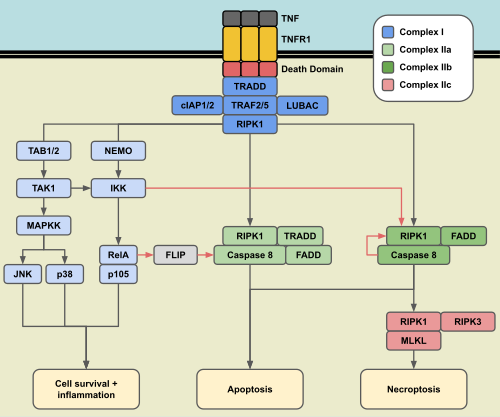
By default, TNFR1 activation triggers Complex I. This occurs when the receptor trimerizes and recruits proteins including RIPK1, TRADD, TRAF2, cIAP1, cIAP2, and LUBAC. Through a process of ubiquitination, these proteins recruit TAK1 and IKK, which activate the MAPK and canonical NF-κB pathways. These pathways trigger transcription factors in the nucleus that promote cell survival and inflammatory responses.
Complex I is regulated by deubiquitinases like A20, CYLD, and OTULIN, which act to destabilize the complex.
Complex II: The Pathways to Cell Death
When RIPK1 or TRADD dissociates from Complex I and binds with FADD, Complex II is formed, activating caspase 8. There are two main variations: Complex IIa (includes TRADD) and Complex IIb (dependent on RIPK1).
To prevent accidental cell death, the body employs three critical cell death checkpoints:
- The IKK Checkpoint: IKK phosphorylates RIPK1 while it is in Complex I, disabling Complex IIb. If ubiquitination is inhibited, this checkpoint fails, leading to apoptosis or pyroptosis (via GSDMD cleavage).
- The NF-κB Checkpoint: The NF-κB pathway expresses pro-survival genes like FLIP, which block caspase 8 in Complex IIa. Translation inhibitors like cycloheximide can disable this checkpoint.
- The Caspase 8 Checkpoint: A non-lethal form of caspase 8 cleaves RIPK1, preventing the formation of the necrosome (Complex IIc). If caspase 8 is inactivated, RIPK1 binds to RIPK3 and MLKL, triggering necroptosis.
TNFR2 Signaling: Proliferation and Cross-talk
TNFR2 is expressed in limited cell types, such as fibroblasts and subsets of immune cells. Unlike TNFR1, it lacks a death domain and cannot directly induce cell death. It is primarily activated by tmTNF.
TNFR2 signaling often leads to cell proliferation via the non-canonical NF-κB pathway. This process requires the accumulation of NIK, which activates IKKα, allowing p100 and RelB to form a heterodimer. Notably, this process is often potentiated by TNFR1's canonical NF-κB activation.
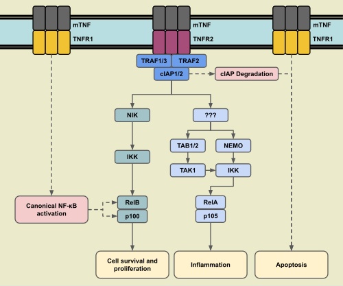
Although TNFR2 generally promotes survival, it can indirectly cause cell death by degrading cIAP1/2. This degradation disrupts the ubiquitination of TNFR1's Complex I, disabling the IKK checkpoint and triggering TNFR1-mediated cell death.
Reverse Signaling and Systemic Immune Response
Transmembrane TNF (tmTNF) can function as a receptor itself, a process known as reverse signaling. The outcome depends on the cell type:
- B lymphoma cells: Increases NF-κB activity, enhancing survival.
- Natural Killer (NK) cells: Increases cytotoxic activity (perforin, granzyme B).
- T cells: Activates the JNK pathway, leading to apoptosis.
- Monocytes: Modulates inflammatory responses to sTNF and endotoxins via TGF-β production.
The Role of TNF in Acute Inflammation
TNF is a primary regulator of acute inflammation. It stimulates endothelial cells to induce coagulation (limiting microbe spread) and secretes chemokines to attract white blood cells. It also signals the liver to produce acute phase proteins, such as C-reactive protein, and can induce muscle and fat catabolism for energy. However, excessive TNF levels can lead to life-threatening septic shock.
Induction of Fever
TNF induces fever by triggering the release of IL-1 and IL-6 or through mediators like PLA2. These signals reach the hypothalamus, leading to the synthesis of prostaglandins, which raise the body's target temperature.
TNF in the Central Nervous System
In the brain, TNF is expressed by neurons and glial cells (microglia and astrocytes) to maintain homeostasis and modulate synaptic plasticity:
- Synaptic Scaling: TNF increases the expression of excitatory AMPA and NMDA receptors while decreasing inhibitory GABAA receptors.
- Neurotransmission: It modulates the release of glutamate and S100B from astrocytes.
- Myelination: Through TNFR2, TNF promotes the maturation of oligodendrocytes, though it can become cytotoxic to their progenitors when in contact with astrocytes.
| Feature | TNFR1 | TNFR2 |
|---|---|---|
| Cell Distribution | Most cell types | Limited (Endothelial, Fibroblasts, Neurons) |
| Ligand Binding | sTNF and tmTNF | Primarily tmTNF |
| Death Domain | Present | Absent |
| Primary Outcomes | Survival, Inflammation, Apoptosis, Necroptosis | Survival, Proliferation, Inflammation |
| NF-κB Pathway | Canonical | Non-canonical (primarily) |
Frequently Asked Questions
What is the difference between apoptosis and necroptosis?
Apoptosis is a controlled, programmed form of cell death. Necroptosis is a less controlled form of death that causes inflammation and can interfere with surrounding tissues.
How does TNF induce fever?
TNF triggers the release of cytokines like IL-1 and IL-6 or uses mediators like PLA2 to signal the hypothalamus. This results in the production of prostaglandins, which increase the body's temperature set point.
Can TNFR2 cause cell death?
TNFR2 cannot directly induce cell death because it lacks a death domain. However, it can indirectly cause death by degrading cIAP1/2, which disables the survival checkpoints of TNFR1.
What is tmTNF reverse signaling?
Reverse signaling occurs when transmembrane TNF (tmTNF) acts as a receptor rather than a ligand, triggering intracellular responses within the cell that expresses the TNF molecule.
Why do pathogens target inflammatory pathways?
Pathogens disrupt inflammatory pathways to avoid an immune response. However, because TNFR1's cell death pathways are inhibited by these same inflammatory signals, disrupting them often triggers the cell to die, preventing the pathogen from replicating.